

Proactive Vision Care at Stonebridge Eyecare
Many serious eye conditions develop gradually without early discomfort or obvious visual changes. Regular comprehensive eye exams play a critical role in detecting potential issues before they permanently alter your sight.
At Stonebridge Eyecare, our team provides thorough assessments, ongoing monitoring, and personalized management for a wide spectrum of eye diseases to support your visual health. We use modern diagnostic technology to make your experience more comfortable. Book your comprehensive exam today.
Using Technology for Early Detection
Eye diseases often develop subtly, advancing quietly beneath the surface without causing pain or sudden visual disruption in their initial stages. Advanced clinical technology allows us to evaluate internal structures and identify early warning signs that might otherwise go unnoticed.
During routine eye care visits, our clinic uses Optomap retinal imaging, OCT scans, and FDT Matrix visual field testing. These diagnostic tools help us check for signs of common conditions, including glaucoma, age-related macular degeneration (AMD), and diabetic eye disease, allowing for timely management and care.

Glaucoma
Glaucoma is a group of progressive eye conditions that damage the optic nerve. This nerve is responsible for sending visual information from your eye to your brain. Glaucoma is frequently associated with elevated pressure inside the eye, though it can occur even with normal intraocular pressure (IOP) readings.
Because glaucoma typically diminishes side (peripheral) vision first, significant damage can accumulate long before you notice changes in your central sight, making it a leading cause of irreversible blindness.
Open-angle glaucoma is the most common form, developing slowly over time as drainage channels in the eye become gradually clogged.
Angle-closure glaucoma occurs when the iris bulges forward, blocking drainage channels suddenly and causing a rapid, severe rise in eye pressure that requires emergency care.
Normal-tension glaucoma happens when optic nerve damage occurs despite eye pressure remaining within normal statistical ranges.
Testing for glaucoma involves a combination of specialized assessments to evaluate both eye pressure and structural nerve health.
Tonometry measures the fluid pressure inside your eye, while optical coherence tomography (OCT) creates high-resolution cross-sectional images of the optic nerve fibres. Additionally, visual field testing, such as the FDT Matrix, evaluates your peripheral vision to detect tiny patches of vision loss.
While damage caused by glaucoma cannot be reversed, ongoing treatment helps stabilize eye pressure to prevent further optic nerve loss. Management typically begins with daily prescription eye drops that lower fluid production or improve drainage inside the eye.
In some cases, oral medications, laser therapy, or surgical intervention may be recommended by a specialist to maintain stable intraocular pressure over time.
Cataracts
A cataract is a gradual clouding of the eye’s natural lens, which sits behind the iris and pupil. As proteins in the lens clump together over time, light struggles to pass cleanly to the retina, causing images to appear blurry or hazy.
Cataracts gradually alter your visual clarity, distort colour perception, and increase sensitivity to glare, particularly when driving at night.
Common symptoms include:
- Cloudy, dim, or double vision
- Seeing faded or yellowed colours
- Increased sensitivity to bright lights
- Halos or ghost images
- Difficulty seeing at night
Aging is the primary risk factor for cataracts, as natural lens proteins change over time.
Extended exposure to ultraviolet (UV) sunlight, diabetes, smoking, and excessive alcohol consumption can also contribute to earlier or faster cataract formation.
Furthermore, prolonged use of corticosteroid medications or previous eye injuries can increase your likelihood of developing cataracts.
In the early stages, cataracts can be managed by updating your prescription eyeglasses and using brighter lighting for reading and daily tasks. As the condition advances and interferes with your routine activities, cataract surgery provides a reliable solution.
During surgery, an ophthalmologist replaces the cloudy natural lens with a clear artificial intraocular lens to restore visual clarity. We provide preoperative guidance and postoperative care.
Age-Related Macular Degeneration
Age-related macular degeneration (AMD) is a condition that affects the macula, the small central portion of the retina responsible for sharp, straight-ahead vision.
As AMD progresses, it impairs your ability to recognize faces, read fine print, drive, or see small details. Individuals experiencing AMD may notice that central vision becomes blurry, dark spots appear in their line of sight, or straight lines look distorted or wavy.
Dry AMD is the more common form, characterized by the gradual thinning of macular tissue and the buildup of yellow cellular deposits called drusen.
Wet AMD is a less common but more severe progression where abnormal, fragile blood vessels grow beneath the retina and leak fluid or blood into the macula. Wet AMD often causes rapid, significant central vision changes that require urgent medical attention.
Age is the strongest risk factor for macular degeneration, with the condition occurring most frequently in individuals over 50.
Family history and genetics play a major role, as does lifestyle; smoking significantly increases your risk of developing AMD.
Uncontrolled high blood pressure, elevated cholesterol levels, and poor nutrition also contribute to the onset and progression of macular changes.
Testing for AMD begins during a comprehensive eye exam using retinal imaging and OCT scans to inspect the macular layers for drusen or fluid.
An Amsler grid test is also used to check for distorted grid lines or blank spots in your central field of vision. These diagnostic tools allow us to identify early structural changes and monitor any subtle shifts in macular health over time.
Management strategies depend heavily on whether you have dry or wet AMD.
For dry AMD, custom nutritional supplementation based on the AREDS2 formula and targeted therapies like MacuMira can support retinal cell health and help preserve vision.
For wet AMD, rapid referral for anti-VEGF injections or laser procedures helps seal leaking blood vessels and manage swelling.
Flashes and Floaters
Flashes and floaters are common visual phenomena related to changes within the vitreous, the gel-like substance that fills the inside of your eye.
While occasional floaters are normal for many people, a sudden influx or change in how they appear can signal an underlying retinal issue. Recognizing these symptoms helps you know when to seek a prompt evaluation.
Flashes appear as quick streaks, arcs, or bursts of light across your field of vision, similar to lightning or camera flashes. They occur when the vitreous gel tugs, pulls, or rubs against the light-sensitive retina inside your eye.
While flashes can be harmless, a sudden increase in flash frequency often indicates retinal traction that needs to be assessed.
Floaters look like tiny specks, threads, cobwebs, or dark spots that drift slowly across your vision when you move your eyes. They are caused by microscopic protein fibres within the aging vitreous casting tiny shadows onto your retina.
Most floaters are harmless signs of aging, but a sudden shower of new floaters can be a warning sign of a retinal tear.
You should seek prompt eye care if you notice a sudden increase in floaters, frequent light flashes, or a dark shadow pulling across your side vision.
These red-flag symptoms can indicate a retinal tear or detachment, which requires urgent treatment to protect your vision. If you experience these changes, contact our clinic immediately for an urgent assessment.
Conjunctivitis
Conjunctivitis, commonly known as pink eye, is an inflammation or infection of the transparent membrane (conjunctiva) that lines your eyelid and covers the white part of your eyeball.
It causes small blood vessels in the eye to become inflamed, resulting in a reddish or pink appearance, irritation, and discharge. Because conjunctivitis can stem from several distinct causes, an accurate clinical diagnosis is necessary to treat the condition effectively.
Allergic conjunctivitis occurs when the eyes react to environmental triggers like pollen, pet dander, dust mites, or mold. It typically affects both eyes simultaneously, causing intense itching, redness, tearing, and puffy eyelids without being contagious.
Management focuses on identifying allergens, using cool compresses, and applying lubricating or antihistamine eye drops.
Viral conjunctivitis is highly contagious and usually accompanies common respiratory viruses or colds. It typically causes watery discharge, redness, a gritty feeling, and light sensitivity, often starting in one eye before spreading to the other.
Because antibiotics do not treat viruses, management centers on supportive care, cool compresses, artificial tears, and strict hygiene to prevent spreading the infection.
Bacterial conjunctivitis is caused by bacteria and is characterized by thick, yellow or green sticky discharge that can cause the eyelids to stick together upon waking. It can affect one or both eyes and is easily spread through direct contact or shared personal items.
Treatment typically involves prescription antibiotic eye drops or ointments to clear the infection and relieve discomfort promptly.
Protect Your Vision with Routine Care
Eye diseases can impact your clarity and long-term visual health, but many can be effectively managed when caught early. From glaucoma and cataracts to AMD, consistent clinical monitoring provides a strong defence against progressive vision loss.
We recommend that everyone schedule a comprehensive eye exam every 1 to 2 years, or as directed by your optometrist, to stay proactive about their eye health.
Frequently Asked Questions About Eye Diseases
Common symptoms include persistent blurriness, dark spots, floaters, sudden flashes of light, or loss of peripheral vision. If you notice any of these warning signs or if it has been more than 2 years since your last exam, please book an eye exam with our clinic in Saskatoon.
Optometrists use digital imaging, pupil dilation, and microscopic evaluations to examine the delicate blood vessels and structures inside your eye. These detailed diagnostic tools allow them to identify subtle signs of eye disease long before noticeable visual changes occur.
Most adults should schedule a comprehensive eye checkup every 1 to 2 years, though individuals with conditions like diabetes may require more frequent visits. If it has been more than 2 years since your last exam, we encourage you to book an appointment with our team today.
No, a physician’s referral is not required to schedule a comprehensive eye exam or medical eye assessment with an optometrist in Saskatchewan. You can contact our clinic directly to book an appointment whenever you have visual concerns or need a routine vision exam.
Yes, progressive conditions like diabetic retinopathy and glaucoma frequently develop silently without causing early pain or noticeable visual loss. Regular comprehensive eye exams are essential to detect these silent conditions early and protect your long-term sight.
Your optometrist will explain the findings, discuss personalized treatment or monitoring strategies, and co-manage your care with specialists if advanced intervention is needed. We will establish an ongoing follow-up schedule to monitor your eye health and help maintain your vision.
Saskatchewan Health Services covers annual eye health assessments for residents diagnosed with diabetes, as well as visits for ocular emergencies. If you notice new visual symptoms or if it has been more than 2 years since your last exam, call our clinic or book an appointment today.

Visit Our Location
We’re located in Stonebridge, on the corner of Cope Lane and Stonebridge Boulevard. Find us next to the Saskatoon Dermatology Centre and Stonebridge Dental Centre. Free parking is available.
Our Address
Contact Information
- Phone: 306-664-2638
- Fax: 306-653-5822
- Email: secadmin@yxevision.ca
After Hours Emergencies: 306-371-9911
Hours of Operation
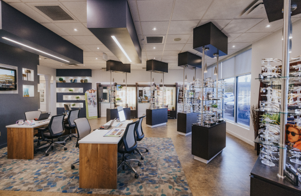
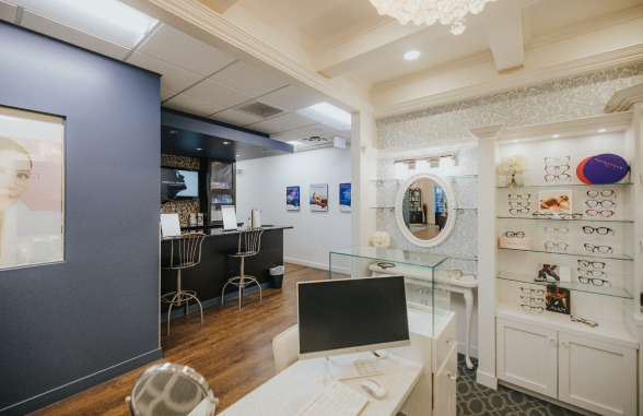
A Look
Inside Stonebridge Eyecare



See What Our Patients Are Saying
